11.005 BF179
Lichen Case-bearer Dahlica lichenella
(Linnaeus, 1761)
Description by Ian F. Smith:
Winged males of Dahlica lichenella occur in Europe, though in Switzerland populations with both sexes are far fewer than those with only the parthenogenetic wingless female form. Only females have been recorded in Britain; in a few places in southern England, Cheshire, southern Scotland and Perthshire, but the species may have been under-recorded because of identification problems, and males might yet be found.
The adult lives a very short time, so the cased larva is more likely to be found on sunny rocks, stone walls, wooden posts and tree trunks. The case, triangular in cross section and tapered at both ends, is covered in granules of sand, lichen and/or algae.
- Description: Ian F. Smith:
The larva grows from 1mm on hatching in May to about 5 mm in late September, overwintering full grown until the following February. The larva pupates in the case, and in March or April the pupa extrudes from the case for the wingless female to emerge and lay her eggs in the vacated case. Positive identification depends on dissection of the genitalia, or examination of the headplate of the pupal exuviae. Specimens should be taken sparingly as excessive collecting could cause localised eradication of this species with limited dispersal ability. Apparently vacated cases with extruded pupal exuviae may contain over 50 ova.
Parthenogenetic female imago: The specimen described was found in mid April in sunshine at 1p.m. on the vacated larval case, which had an extruded pupal exuviae. It oviposited into the case until 5pm, when it became inactive, and died during the night. Length: 4 mm (3mm when ovipositor retracted). Head: Pitchy black. Antenna filiform with 16 segments (MBGBI, 14-19 segments). Thorax: Dorsum, sides and thoracic legs glossy brown. 4 segments on tarsus. Wings, vestigial whitish flaps. Abdomen Segments A1-A7: Dorsum glossy brown, as thorax, or a bit paler in some lighting. Each segment with a whitish posterior edge which protrudes at times. Laterally whitish with dense cover of backwardly swept dark brown-golden hairs. Venter glossy brown, slightly lighter than dorsum. A skein of white silk bent into a 'v' on either side of venter of segment A7. The silk may become detached and inserted with the eggs into the case. Segment A8: Modified into a pitchy black extendible ovipositor.
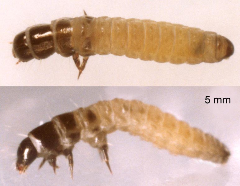
Larva:
Food: Lichen and algae on rocks, stone walls, wooden posts and tree trunks in sunny situations. MBGBI indicates larvae from June to March, but observations in Cheshire suggest May to February. In the Alps, pupation is probably at the time of snow melt as the males fly a month later in early March to mid-May, depending on the date of melt at different altitudes.
First instar Length: 1 mm (May in Cheshire) Case: 1 mm. Silk coated with fine sand, algae and lichen granules. Head: Yellowish brown. Stemmatal area dark chestnut. Prothorax (T1): Prothoracic shield yellowish brown. Mesothorax (T2): Translucent white showing yellowish brown of viscera in posterior half. Metathorax (T3): As T2, but yellowish brown less strong. Thoracic legs: Transparent light brown. Body: Yellowish white. Spiracles: Concolorous, inconspicuous. Abdominal pinacula: Concolorous. Setae: Colourless transparent. Anal plate: Light brown. Prolegs: Concolorous. Crochets reddish brown. Ventral prolegs small.
Late instar (Probably final instar) Length: 5 mm (September, larva extended and walking, Cheshire). 4 mm (January, larva contracted and awake but resting, Cheshire). Case: 6 or 6.5 mm, Cheshire (MBGBI; 5-7 mm). Triangular cross section, tapered both ends. White silk covered in sand, lichen and algae. Pupa 4 mm, extruded from end in March or April. Head: Black. Mouthparts reddish brown. Fine colourless epicranial and adfrontal cleavage lines. Prothorax (T1): Large pitchy black prothoracic shield, divided by thin transparent colourless medial line. Shield covers entire segment dorsally and laterally, apart from white membrane visible at anterior when head extended. Ventrally and intersegmentally, greyish white integument. Mesothorax (T2): Dorsum covered by large pitchy brown sclerite, divided by transparent colourless medial line which is widest at the posterior, and overall wider than the dividing line on T1. Large greyish brown lateral sclerite and subventral sclerite. Integument between sclerites greyish white. Metathorax (T3): Colourless transparent subdorsal sclerite at anterior. Large triangular dorsolateral sclerite, pitchy at centre fading to greyish brown at edges. Whitish grey lateral sclerite and subventral sclerite. Thoracic legs: Dark brown with thin black lines on femur. Base of legs coloured as venter. Leg on T3 markedly longer, due mainly to a very swollen base. When standing, the long legs on T3 cause the body to form a characteristic attenuated arch. At rest, the legs on T3 are inclined backwards or forwards, and the body is shorter and less attenuated. Body: Matt greyish yellow in September, whitish ochre in January. Spiracles: Small and inconspicuous with fine brown peritreme. Abdominal pinacula: Segments A1-A9 each have a subdorsal, dorsolateral and lateral sclerite, shiny and slightly darker than the integument . Setae: Fine, colourless transparent on abdomen. Brown on thorax. Anal plate: Large, covering all dorsum of A10. Greyish centrally, blackish laterally. Prolegs: On A3-A6, vestigial and concolorous with venter. On A10 proleg has swollen greyish base. Crochets reddish brown. When extracted from case and standing, the only prolegs in contact with the substrate are those on A10, and sometimes the posterior rests on the bent-under anal plate instead of on them.Similar species: There has been much confusion with the identification of Dahlica spp. Useful references are MBGBI 2 and Schmetterlinge und ihre Lebensräume, Band 2. Published by Schweizerischer Bund für Naturschutz. (The Psychid contributions in both are by P. Hättenschwiler.) The female pupal headplate, often found displaced but still attached to the pupal exuviae, is a useful diagnostic feature. However, care should be taken, as the individual elements are rarely disposed in exactly the same positions after emergence. The angle of view can also cause variation.
- 175 Narycia monilifera has a similar case and larva. The sclerites on the larval thorax are very similar, except that the dorso-lateral sclerite on T3 is smaller and rounded on N. monilifera, and larger and triangular on Dahlica lichenella. The abdomen of N. monilifera is partly translucent showing viscera medially on the dorsum or more extensively, while that of D. lichenella is opaque. Shiny sclerites, slightly darker than the integument, are discernible on the abdomen of D. lichenella, but absent from N. monilifera. The abdomen of N. monilifera is usually too plump for the larva to be expelled from the case without slitting it open, while D. lichenella is slim enough to be expelled by progressive careful pressure on the case starting from the rear. Both sexes of Narycia are winged, and the female flies to distribute her ova on different tree trunks. Consequently her ova are not found in the vacated larval case, and the cased larvae are usually found scattered on different trunks in ones or twos, not concentrated in groups of twenty to a hundred or more, as with D. lichenella. In late March, April and May, triangular cases with full grown larvae are likely to be N. monilifera as, in Britain, all three Dahlica species will have pupated by then, unless arrested by parasitoids, and their new larvae will be only 1 or 2mm long.
- 185 Luffia ferchaultella, the parthenogenetic form of 184 L. lapidella, also occurs in localised concentrations of larvae, but its larval case is rounded in cross section. 'Schmetterlinge und ihre Lebensräume, Band 2' Published by Schweizerischer Bund für Naturschutz states that larvae of the species in the genus Dahlica are very similar. The pupal headplate, often found displaced but still attached to the pupal exuviae, is a useful diagnostic feature. However, care should be taken, as the individual elements are rarely disposed in exactly the same positions after emergence. The angle of view can also cause variation.
- 176 Dahlica triquetrella: Only wingless females in Britain. (Males occur at some mainland Europe sites). Case has fragments of insect attached. (Beware insect fragments on 180 Diplodoma herminata and anterior 20% of 181 Taleporia tubulosa cases.) Pupal headplate has base of antenna slightly swollen. (MBGBI; neck section not indented) The adult has 5 segments in the tarsus of the foreleg ( Schweizerischer Bund für Naturschutz).
- 177 Dahlica inconspicuella: Wingless females and winged males. Not known outside Britain. Case coated with lichen or algae. Pupal headplate has base of antenna swollen.
- 179 Dahlica lichenella: Only wingless females in Britain. (Males occur at some mainland Europe sites). Case coated with sand and lichen or algae. Pupal headplate has base of antenna slender. (MBGBI; neck section of headplate indented by antenna base. Note that this does not show in the views on this website or in Schmetterlinge und ihre Lebensräume, Band 2.) The adult has 4 segments in the tarsus of the foreleg (Schweizerischer Bund für Naturschutz).
More images